Articles
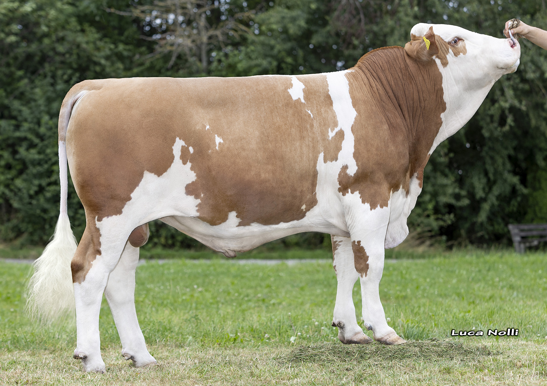
Broad CRV Fleckvieh range offers top-quality bulls for every breeding goal
Broad CRV Fleckvieh range offers top-quality bulls for every breeding goal Article and photo courtesy of CRV All Fleckvieh breeders have their own personal requirements. CRV’s Fleckvieh program offers a broad range of bulls that will always meet ones breeding goal, whether that is on kilos milk, on increasing fat and protein or on achieving […]
READ MORE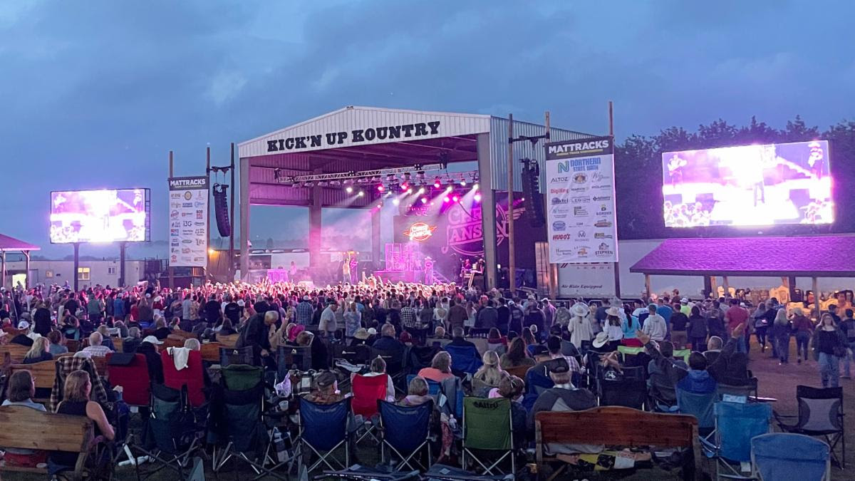
Mattracks Partners With Kick’n Up Kountry For 21st Year
Mattracks Partners With Kick’n Up Kountry For 21st Year Article and photo courtesy of Mattracks Karlstad, MN – April 23, 2024 – Mattracks has once again joined forces with Kick’n Up Kountry (KUK) in preparation for its highly anticipated 2024 music festival. Kick’n Up Kountry, which takes place annually at its permanent home at Wagon […]
READ MORE
Can high cattle prices pay for mistakes?
Can high cattle prices pay for mistakes? Article and photo courtesy of MU College of Agriculture, Food and Natural Resources. SALEM, Mo. – With cattle prices at record high levels and many forecasts projecting relatively high prices for the next couple of years, some producers feel they can do no wrong. In times like these, […]
READ MORE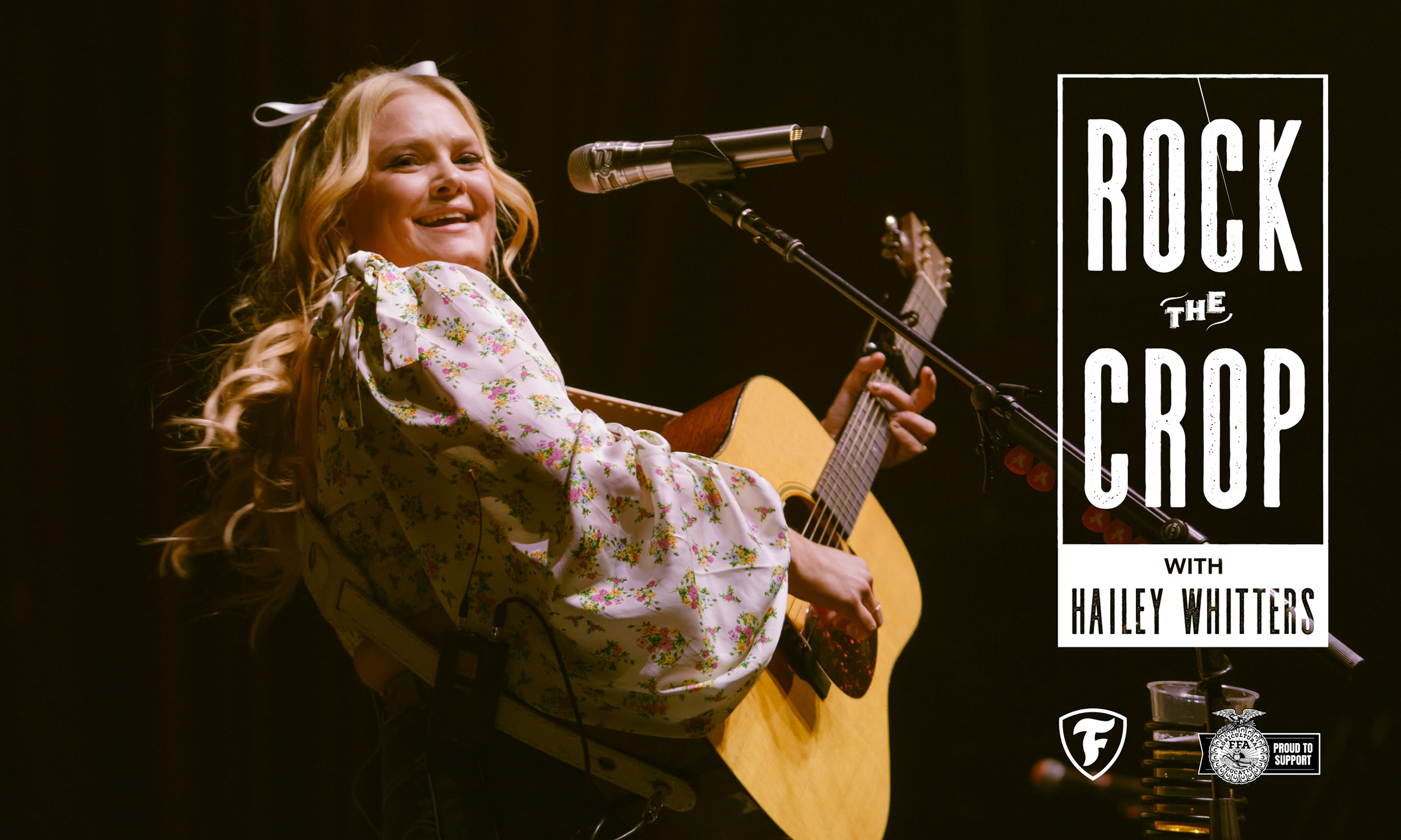
Firestone Ag Opens 2024 Rock the Crop Sweepstakes Exclusively for National FFA Chapters
Firestone Ag Opens 2024 Rock the Crop Sweepstakes Exclusively for National FFA Chapters Article and photo courtesy of Firestone Ag NASHVILLE, Tenn. (April 24, 2024) – Firestone Ag, a business of Bridgestone Americas (Bridgestone), has opened entries for its 2024 Rock the Crop Sweepstakes exclusively to chapters of the National FFA Organization (FFA) in celebration […]
READ MORE